Retained foreign bodies are uncommon but recognized complications of invasive medical procedures, including those considered minimally invasive. In a recent case series, Hamed Zartab, MD, of Tehran University of Medical Sciences, and colleagues described two patients with retained metallic fragments in the facial region that were identified months following cosmetic and dental procedures. The cases demonstrated delayed presentation and the role of imaging in diagnosis.
Retained surgical foreign bodies may present immediately following a procedure or remain undetected for months or years, according to the researchers. Clinical manifestations may include pain, swelling, discharge, or firm palpable findings, although symptoms may be mild or absent. Imaging modalities used to identify retained foreign bodies include plain radiography, ultrasonography, computed tomography, and magnetic resonance imaging, the researchers reported.
The first case involved a 42-year-old woman who presented 10 months following submental liposuction and lipotransfer performed for aesthetic purposes. She reported a firm, band-like sensation beneath her chin with intermittent mild discomfort. Physical examination revealed a firm, palpable structure in the submental region without overlying skin changes, signs of infection, limitation in neck movement, or discomfort with swallowing.
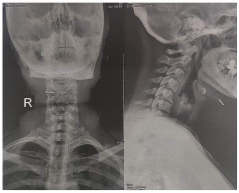
Based on the clinical presentation and procedural history, the differential diagnosis included postoperative fibrosis, fat necrosis, calcified hematoma, granuloma, and retained foreign body related to the prior liposuction procedure. Neck radiography demonstrated an opaque, linear structure located anterior to the hyoid bone on both anteroposterior and lateral views.
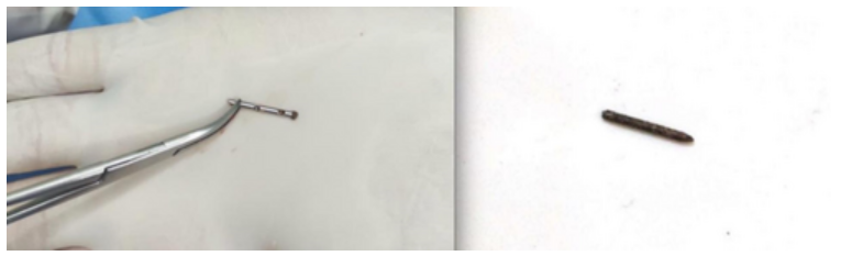
Under local anesthesia, clinicians surgically explored the area through a small incision and removed the retained object. The foreign body was identified as a fractured liposuction cannula tip measuring approximately 2 centimeters in length. The patient was prescribed oral cephalexin 500 mg every 8 hours. Weekly follow-up visits were completed for 2 weeks. The researchers reported that the patient recovered uneventfully and that her symptoms resolved completely during follow-up. Written informed consent was obtained.
The second case involved a 65-year-old woman who presented with an asymptomatic, firm nodule on the left cheek that had persisted for 5 months. The lesion was located approximately 2 centimeters lateral to the oral commissure. The patient reported undergoing a dental procedure on the same side of the face approximately 2 months prior to the development of the nodule. Physical examination revealed a non-tender, firm nodule measuring approximately 10 by 20 millimeters.
The differential diagnosis included post-traumatic fibrosis, foreign body granuloma, injection granuloma, epidermal inclusion cyst, and retained metallic fragments from dental instruments. Ultrasonography revealed a dermal collection measuring 17 by 8 by 6 millimeters with a central echogenic linear structure measuring approximately 10 millimeters, findings suggestive of a retained foreign body.
Surgical exploration was performed under local anesthesia with lidocaine. An incision was made at the point closest to the skin surface, and the foreign body was extracted using forceps. The object was identified as a retained dental needle. The patient attended follow-up visits at the end of the first and second weeks and recovered without complications, according to the researchers. Written informed consent was obtained.
In the discussion, the researchers noted that retained foreign bodies may occur following minimally invasive procedures, including aesthetic and dental interventions. They reported that stainless steel foreign bodies may remain relatively inert, which can delay inflammatory responses and complicate early diagnosis. Plain radiography was described as a reliable imaging modality for detecting metallic foreign bodies in soft tissues, while ultrasonography was effective in identifying retained metallic objects without radiation exposure.
The researchers highlighted that early detection and removal of retained foreign bodies are important to prevent complications, including fibrosis, infection, migration, or inflammation. They also highlighted the importance of careful inspection of surgical instruments before and following invasive procedures and timely communication with patients if instrument failure occurs.
The researchers reported no conflicts of interest. No external funding was reported. Written informed consent was obtained from both patients included in the case series.
Images reproduced from Zartab et al., Clinical Case Reports (2026), licensed under CC BY-NC-ND.
Source: Clinical Case Reports