With its fully automated function, compact design, and expandable modules and features, Topcon Healthcare’s Maestro was well received by the market when it was first launched in 2014 due to its user-friendly features. With the advent of OCT angiography (OCT-A) and its ability to present the vascular network of the retina, and the one page Hood glaucoma report, Topcon launched the Maestro2 in 2018, which quickly became the leading product in the company’s OCT portfolio. Global sales topped 10,000 units in 2019 and rose to 15,000 in 2021. In March 2023, the Maestro2 is approaching its next sales milestone of 20,000 units worldwide.* In the UK, the device is also approved for use on the NHS Diabetic Eye Screening (DES) Program, an annual screening service to check for eye problems caused by diabetes.
The rapid adoption of the Maestro2 – a fully automated, compact system that combines spectral domain OCT, a non-mydriatic high-resolution retinal camera for true color fundus photography, and OCTA** – has been propelled by its broad clinical applications and benefits. The device is helping clinicians meet the challenges of an ever-increasing glaucoma and retinal disease treatment workload, allowing them to handle a large volume of patients and quickly assimilate information in each case. Users have praised the Maestro2 for its intuitive simplicity, efficiency, and ability to provide a comprehensive range of detailed reports to assist practitioners in clinical care. It features excellent image quality and advanced diagnostic capabilities, while offering workflow enhancements, detailed image analysis and reporting functions, and data management capabilities.
Clinical utilities
The Maestro2 is extremely easy to use – allowing less experienced operators to capture quality images with minimal training – and also offers a wide range of clinical utilities. IMAGEnet 6 capture software enables dynamic viewing of OCT and imaging data. Topcon’s PinPoint Registration precisely matches specified areas within OCT and OCTA scans upon the color fundus image. The follow-up scan feature scans the exact same location each patient visit, beneficial for follow-up visits and tracking diseases over time. And an extensive portfolio of reports for macula, anterior, and glaucoma, including a dedicated one-page glaucoma report designed by Professor Donald Hood, allows the practitioner access to advanced diagnostic data (see Table).
The Maestro2’s other capabilities include
- 12 x 9 mm scanning and seven-layer automated segmentation, encompassing both the macula and optic disc, ideal for an annual eye exam and reducing patient testing time. It provides thickness and reference data for the retina, RNFL, and ganglion cell layers together with a glaucoma report, which includes disc topography.
- Instantaneous vascular flow information at the touch of a button -- without the need for contrast dye injection -- together with comprehensive segmentation to enable advanced diagnosis.
- Automatic alignment, focus, optimization, and capture. After image capture, the report can be immediately displayed by clicking on the icon.
- Option to switch to manual control for patients who have conditions which make imaging challenging
- Easy connectivity – all reports can be printed or exported in common file formats to Topcon Healthcare’s Harmony cloud-based data management system and other image management systems and electronic medical records.
- Follow-up support for clinical assessment of treatment response or disease progression.
- Anterior segment imaging using the optional anterior headrest support.
Practical and efficient
In designing the Maestro2, Topcon Healthcare’s engineers drew on knowledge from eye care professionals and end-users around the world to inform the development of a complete clinical workstation that would not only enable workflow efficiency, but also fit into small consulting rooms where space is at a premium. The touchscreen monitor rotates a full 360°, allowing operators to distance themselves from the patient or position the device in tight space if needed. This small footprint, combined with the ability to provide valuable information with a single scan, emphasizes the device’s overall user-friendliness and effectiveness. In short, the Maestro2 is a comprehensive OCT system and an indispensable tool in clinical practice.
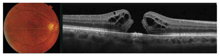
![Figure 2: Color fundus image and OCT scan from an 85-year-old male with retinal vein occlusion.]](https://vision-umbraco.euwest01.umbraco.io/media/msrm0hor/0323-900-topcon-dsf-inarticle-fig2.png)
*Release dates may vary depending on the region. OCTA and Anterior Segment OCT are optional extra features in some countries.
**20,000 units since the Maestro was first launched, inclusive of Maestro2 sales figures.
Not all products, services or offers are approved or offered in every market and products vary from one country to another. Contact your local distributor for country-specific information.
Figures and Images

0323-900-Topcon-DSF-InArticle-FIG1.png