The field of digital dentistry is undergoing a major transformation that is being driven by advancements that enhance precision, streamline workflows, and ultimately, improve patient outcomes. Among the most recent innovations in full-arch implant rehabilitation is intraoral photogrammetry, which is a cutting-edge technology that eliminates many of the limitations associated with traditional intraoral scanning methods.1 Although intraoral scanners have long been the standard for digital impressions, they can struggle with cumulative errors and distortions when capturing multiple implants in full-arch cases.2 The use of extraoral photogrammetry can improve accuracy in full-arch cases; however, these systems still present drawbacks and are often priced beyond the reach of many dentists.
For decades, clinicians have faced challenges in accurately capturing the spatial relationships of multiple implants. Traditional intraoral scanning relies on continuous image acquisition and stitching algorithms, which can introduce distortions when scanning across a large area, such as a full arch with multiple implants.3 Even small alignment errors between individual scans can lead to cumulative inaccuracies, and these inaccuracies can affect the fit of the final prosthesis and require extensive chairside adjustments. In addition, traditional intraoral scanning systems struggle to capture soft-tissue topography to maintain the needed fidelity for precise prosthetic intimacy. These limitations have made full-arch implant rehabilitation one of the most technically demanding procedures in dentistry.4 Intraoral photogrammetry overcomes these challenges by using coded scan markers that ensure precise implant positioning without relying on image stitching.
A Paradigm Shift in Total Record Accuracy
With intraoral photogrammetry technology, clinicians now have access to a highly accurate, efficient, and streamlined workflow for full-arch implant prosthetic planning. Unlike traditional intraoral scanning, intraoral photogrammetry employs specialized horizontal coded scan bodies (High-Accuracy Coded Scan Body, Shining 3D Dental) that are placed over the implant multi-unit abutments to serve as precise reference points (Figure 1). Instead of relying on sequential image stitching, photogrammetry captures the exact 3D spatial relationships between the implants in a single step. This eliminates cumulative errors and provides a digital representation that is far more precise and reliable than that of conventional scanning techniques.
One of the most transformative aspects of intraoral photogrammetry is its ability to seamlessly transition from scan flags to scan bodies within a unified project file. Previously, clinicians needed to capture multiple separate scans at different stages of the treatment, which had the potential to lead to fragmented data and misalignment. With modern intraoral photogrammetry systems, the workflow is now all-in-one, allowing for easy conversion from high-accuracy horizontal coded scan bodies to users' preferred scan bodies (Figure 2) without requiring additional scans. This feature simplifies the transition from surgery to fabrication of the prosthesis and ensures that relative implant and abutment positions remain consistent through the digital workflow.5
Enhanced Soft-Tissue Matching and Efficiency
Another major advantage of intraoral photogrammetry is its ability to accurately capture soft-tissue contours. Achieving an optimal emergence profile and soft-tissue adaptation is critical for full-arch restorations because the final prosthesis must integrate seamlessly with the surrounding gingival architecture. Traditional intraoral scanners often struggle to maintain accuracy when capturing soft tissue, especially in cases where the tissue dynamics change due to pressure from the scan bodies or movement during scanning. With intraoral photogrammetry, the precise implant positions are recorded without the need for direct soft-tissue alignment, which facilitates improved tissue-matching accuracy in the final restoration.
This soft-tissue precision plays a key role in the design and function of the prosthesis. By incorporating tissue-matching algorithms within the digital workflow, clinicians can ensure that the final restoration mimics the natural gingival contours, which reduces the need for excessive chairside adjustments and improves patient comfort. This is particularly important for all variations of fixed prostheses and implant-supported overdentures, for which soft-tissue adaptation directly impacts esthetics and long-term stability.
The introduction of coded healing caps has further improved the efficiency of this workflow. Traditionally, implant scanning required the removal of healing abutments, the placement of scan bodies, and additional scanning steps before implant position data could be captured. With coded cap scan bodies (Coded Cap Scan Body, Shining 3D Dental), the implant positions are captured directly during the scanning phase, eliminating the need for additional scan body placement (Figure 3). The highly scannable shape, surface, and coding of coded healing caps reduces chair time, minimizes errors, and streamlines the transition from healing to design of the final prosthesis, even in a harsh, bloody soft-tissue environment.
Integrating Facial Scanning for Optimal Esthetics
Although intraoral photogrammetry can help to ensure precision in implant positioning, facial scanning can play an equally important role in achieving natural-looking full-arch restorations.6 Traditional workflows have relied on 2D facial photography and analog facebows to determine midlines, lip support, and smile dynamics. These conventional methods only offer a frontal view, without the ability to oscillate the head for cant and anterior-posterior analysis. This limitation introduces variability and fails to present the full 3D relationship between the proposed prosthesis and the patient's facial anatomy.7
With modern 3D facial scanning, clinicians can digitally map key facial landmarks, including the smile line and midline, as well as assess lip mobility and facial symmetry (Figure 4). One of the greatest advantages of integrating facial scanning is its ability to enhance digital smile design. It allows for real-time visualization of the prosthetic design within the patient's facial structure, ensuring that the restoration aligns naturally with the patient's overall appearance.8
The All-in-One Digital Workflow
To improve digital precision, intraoral scanning, intraoral photogrammetry, and facial scanning can be combined into a comprehensive workflow. The process of merging these datasets involves aligning intraoral scans, intraoral photogrammetry data, and facial scans into a unified digital project file (Figure 5). By using common reference points, such as natural teeth or temporary markers, digital software can accurately register all of the datasets together. This enables simultaneous surgical and prosthetic planning, which ensures that the implant positions, bone structure, soft tissue, and esthetics are all considered in a single digital environment.
When intraoral photogrammetry data is merged with intraoral and facial scans, clinicians can preview the final restoration in 3D simulation software. This allows for adjustments prior to fabrication, which not only improves patient communication and treatment acceptance but also reduces the need for multiple trial prostheses, streamlining the entire workflow. Furthermore, being able to view patients in a 3D profile may offer guidance regarding future tooth and flange positions.
This all-in-one approach is also advantageous in improving multidisciplinary collaboration. Oral surgeons, prosthodontists, and dental technicians can all work from the same digital dataset, ensuring that both the surgical and prosthetic goals are aligned. This helps to reduce surgical complications, minimize prosthetic errors, and facilitate more predictable outcomes. The following case report demonstrates how this integrated digital approach can transform clinical workflows to provide a more efficient and precise solution for complex full-arch restorations.
Case Report
A 57-year-old male patient presented to the clinic with a poor-fitting maxillary partial denture and periodontally compromised remaining dentition. He desired full-mouth rehabilitation to replace his failing teeth but had severe dental anxiety, which had contributed to years of neglect. After discussing treatment options, the patient ultimately chose a same-day treatment approach that allowed for preoperative digital planning, surgery under sedation, and immediate delivery of temporary prostheses. The ability to complete surgery and receive temporary full-arch restorations in a single visit alleviated his primary concerns about the need for multiple surgical appointments and extended treatment timelines.
Preoperative Digital Records and Treatment Planning
To ensure a streamlined workflow, comprehensive preoperative digital records were obtained, including clinical photographs, cone-beam computed tomography (CBCT) scans (CS 9600, Carestream Dental), intraoral scans (Aoralscan Elite, Shining 3D Dental), and facial scans (MetiSmile, Shining 3D Dental) (Figure 6 through Figure 9.) These datasets were then integrated into digital planning software to assess bone availability, gingival health, and the transition zone-all critical factors in determining the most appropriate prosthetic solution. Given the patient's condition, dual-arch FP3 restorations were recommended, which would be delivered using a 3D printed stackable guide system (CombiGuide™, ROE Dental Laboratory) to ensure precise implant positioning and surgical predictability.9
One of the key advantages of incorporating intraoral photogrammetry into the workflow was its ability to match intraoral scans seamlessly within its native software. To enhance this scan-matching capability, reference markers were strategically placed on the stackable surgical guide with coded scan bodies (High-Accuracy Coded Scan Body, Shining 3D Dental) attached to enable efficient alignment of the surgical and preoperative records (Figure 10).
Guided Surgical Workflow With Seamless Scan Integration
Prior to sedation, a bite registration was recorded to ensure accurate occlusion for the final prosthesis. This was a crucial step because capturing an accurate bite registration during surgery can be challenging when the patient is under sedation.
After the patient was fully anesthetized, full-thickness flaps were elevated, and the pin-fixated bases of the stackable surgical guide system were anchored, utilizing the remaining teeth for insertion stability (Figure 11 and Figure 12). The remaining teeth were then scanned in relation to the fixation bases, which served as fiducial references to preserve the bite pre- and postextraction. Following extraction, guided bone reduction was performed (Figure 13), and the implant osteotomies were completed through the stackable guide system to ensure ideal positioning.
After the implants (AnyRidge®, Megagen America) were placed, primary stability was confirmed, and the torque and ISQ values supported immediate loading with multi-unit abutments (Figure 14). With this confirmation, the decision was made to proceed with same-day delivery of the interim prostheses.
Intraoral Photogrammetry for Immediate Conversion
To digitally record the implant positions within the same workflow, high-accuracy coded scan bodies and cap scan bodies were fixed to the multi-unit abutments and captured using the intraoral photogrammetry system (Aoralscan Elite, Shining 3D Dental) (Figure 15 through Figure 18). Unlike conventional intraoral scanning or extraoral photogrammetry, intraoral photogrammetry eliminates cumulative stitching errors, providing a precise and direct capture of the implant positions within a fully digital workflow.
Once the high-accuracy coded scan bodies were captured, they were scan-matched to the fixated bases-a crucial bite-recapture step that simplifies the same-day restoration workflow (Figure 19 through Figure 21). This process would significantly reduce design turnaround time and enable the technician to immediately begin fabrication of the restorations without the need for complex manual alignment or best-fit algorithms.
A key benefit of intraoral photogrammetry over extraoral photogrammetry is its ability to streamline the transition from multi-unit abutment photogrammetry flags to multi-unit abutment scan bodies within the same software workflow. Traditional extraoral photogrammetry workflows often require technicians to manually align scan data or use point-matching algorithms to register implant positions, which leads to additional processing time. In contrast, intraoral photogrammetry's integrated soft-tissue scan matching and direct multi-unit abutment selection feature eliminates these extra steps, making same-day prosthesis design and conversion significantly more efficient.
After the photogrammetry was completed, healing caps were secured, and guided bone regeneration was performed where necessary to optimize long-term implant stability. Given the patient's severe dental anxiety, the need for postoperative treatment was minimized whenever possible to help ensure a comfortable recovery process. The surgical sites were closed using resorbable sutures, further reducing the need for postoperative intervention.
Design and Fabrication of the Provisional Prostheses
Following surgery, the intraoral photogrammetry scans were immediately sent to the laboratory technician (ROE Dental Laboratory) via the software's integrated portal (Shining 3D Dental Cloud, Shining 3D Dental). The preoperative digital design was then utilized to expedite the design of the provisional restorations (Figure 22). This step is significantly accelerated when implant positioning is guided because the primary modifications required are determining the exact locations of the screw access holes and finalizing implant angulation.
The digital files were downloaded and transferred to the nesting software (AccuWare, Shining 3D Dental), where both the upper and lower arches were prepared to be simultaneously printed using temporary crown and bridge resin (Temporary CB Resin [CB 11], Shining 3D Dental). The high-speed printing capabilities of the 3D printer system (AccuFab-CEL, Shining 3D Dental) allowed for dual-arch fabrication in under 15 minutes, and its self-cutting wash system automatically detached the printed provisional restorations from the ceramic build plate and dropped them directly into an alcohol agitating bath for efficient post-processing.
After cleaning, the printing supports were manually removed, and the provisional prostheses were stained and glazed to enhance their esthetics. The automated curing system then finalized the restorations, leveraging WiFi integration to synchronize print, wash, and cure settings without manual input. The intelligent workflow automation ensured that material parameters and curing durations were optimized, eliminating guesswork and manual adjustments from the process.
The entire process, from sending the digital files to completing the printed restorations, was completed in under an hour. This added to the efficiency of the workflow and ensured minimal downtime between surgery and delivery of the prostheses.
Delivery of the Provisional Prostheses and Occlusal Optimization
Per the preoperative plan, the provisional restorations, which were designed for a direct-to-multi-unit abutment screw-retained approach (Vortex™, Louisiana Dental Implant Lab), were inserted with the posterior occlusion slightly out of contact, and the screw access holes were sealed with blue PTFE tape and a temporary light-cure filler. After delivery, postoperative photographs and a panoramic radiograph were acquired to confirm the fit (Figure 23 and Figure 24). The patient was instructed to maintain a soft diet to facilitate implant integration.
One of the key benefits of using this intraoral photogrammetry-based workflow is that it significantly reduces the need for postoperative occlusal adjustments. Although occlusal equilibration is often necessary due to minor scan misalignments, the authors of this article have observed a notable decline in postoperative refinement appointments since adopting this system. In this case, no occlusal adjustments were required immediately after placement, and the patient reported uneventful healing at the 10-day postoperative follow-up visit.
Finalization of the Case
The patient was monitored for 3 months to allow for complete osseointegration before delivery of the definitive restorations. Throughout this period, no concerns were noted by the patient aside from his increasing desire to return to chewing hard foods.
Following the observation period, the patient returned, and final digital records were obtained. This included the acquisition of new high-accuracy intraoral photogrammetry scans to confirm implant positioning and precisely match the provisional restorations to the soft-tissue contours, ensuring an accurate transition to the final prosthesis (Figure 25 through Figure 28). The final zirconia prostheses (Bespoke, Roe Dental Laboratories) were designed based on these updated digital records.
Once fabricated, the final zirconia prostheses were passively inserted direct-to-multi-unit abutment. The only notable refinement to the occlusion that was required involved a minor interference from an upper molar during lateral excursive movements. It was quickly resolved.
Six months postoperatively, the patient returned for a final panoramic radiograph and intraoral and extraoral photographs, all of which confirmed excellent tissue stability and a precise prosthetic fit (Figure 30 through Figure 36). He expressed significant satisfaction with both the function and esthetics of the restorations and highlighted his newfound ability to chew comfortably.
Beyond the clinical success of the case, the patient's dental anxiety dramatically diminished throughout treatment. What began as a highly anxious individual transformed into a confident advocate for digital dentistry who referred multiple new patients for similar treatments. His case serves as a testament to the power of predictable, efficient, and patient-centric digital workflows, reinforcing the value of intraoral photogrammetry, same-day treatment protocols, and cutting-edge restorative technologies in full-arch implant rehabilitation.
Conclusion
This case demonstrates how the combination of stackable surgical guides and intraoral photogrammetry with advanced coded scan bodies can transform full-arch implant rehabilitation into a predictable, efficient, and patient-friendly procedure. By eliminating the inaccuracies of traditional intraoral scanning, intraoral photogrammetry allows clinicians to achieve unprecedented levels of precision. When integrated with facial scanning, this technology ensures that final restorations are both functionally precise and esthetically natural looking, reducing the need for chairside modifications and enhancing patient satisfaction.
With intraoral photogrammetry's advanced scan-matching capabilities, the transition from surgical placement to prosthetic design is seamless, accurate, and significantly faster than traditional workflows. The ability to capture implant positions, match scan flags to the surgical guide, and integrate soft-tissue data within a single project file-and when necessary, seamlessly transition from coded healing caps to final scan bodies-optimizes efficiency, which ultimately reduces chair time and improves the overall patient experience. As the technology continues to evolve, intraoral photogrammetry may become the gold standard in full-arch implant rehabilitation, offering a fully digital, highly predictable, affordable, and patient-friendly solution for modern implant dentistry.
About the Authors
Isaac Tawil, DDS, MS
Founder and Co-director
Advanced Implant Education
Private Practice
Brooklyn, New York
Michael Erdos, DDS
Private Practice
Brooklyn, New York
References
1. Tawil I, Ganz SD, Pozzi A. Intraoral photogrammetry: the next step in full arch implant precision. International Magazine of Digital Dentistry. 2024;4:42.
2. Tawil I, Domingue D. Navigating the complexities of digital full-arch implant treatment: strategies to improve accuracy and deliver optimal outcomes. Inside Dentistry. 2024;20(6):33-40.
3. Tawil I, Ganz SD. Fully digital full arch? Continued advancements in full-arch implant restorations. Dentistry Today. 2023;42(4):50.
4. Mangano FG, Hauschild U, Veronesi G, et al. Trueness and precision of 5 intraoral scanners in the impressions of single and multiple implants: a comparative in vitro study. BMC Oral Health. 2019;19(1):101.
5. Jensen OT, Ross D, Jivraj S, Tawil I. Provisional prosthetic outcome when using photogrammetry for complete arch oral implants: a report of 111 patient treatments. Oral Maxillofac Surg Clin North Am. 2025;37(2):179-192.
6. Pozzi A, Arcuri L, Moy PK. The smiling scan technique: facially driven guided surgery and prosthetics. J Prosthodont Res. 2018;62(4):514-517.
7. Tawil I, Domingue D, Ganz SD. Digital full-arch maxillary rehabilitation. Inside Dentistry. 2024;20(7):28-35.
8. Mai H-N, Win TT, Tong MS, et al. Three-dimensional morphometric analysis of facial units in virtual smiling facial images with different smile expressions. J Adv Prosthodont. 2023;15(1):1-10.
9. Ganz SD, Tawil I. Full-arch implant surgical and restorative considerations: utilizing a full template guidance technique. Dentistry Today. 2019;38(9):72-78.
Figures and Images

Fig. 1

Fig. 2

Fig. 3

Fig. 4

Fig. 5
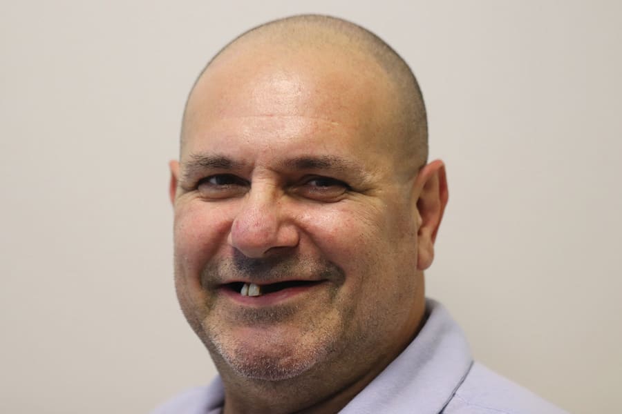
Fig. 6

Fig. 7

Fig. 8

Fig. 9

Fig. 10

Fig. 11

Fig. 12

Fig. 13

Fig. 14

Fig. 15

Fig. 16

Fig. 17

Fig. 18

Fig. 19

Fig. 20

Fig. 21

Fig. 22

Fig. 23

Fig. 24

Fig. 25

Fig. 26

Fig. 27

Fig. 28

Fig. 29

Fig. 30

Fig. 31

Fig. 32

Fig. 33

Fig. 34

Fig. 35

Fig. 36