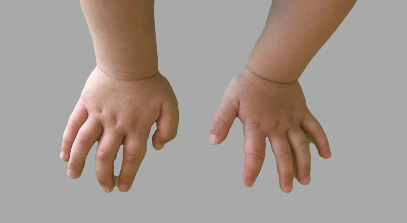
A 3-year-old Italian boy with a history of neurodevelopmental delay, autism spectrum disorder, mild facial dysmorphisms, and congenital anomalies was diagnosed with a de novo 13.4-Mb triplication of chromosome region 3p24.3p23, a previously unreported genetic variant potentially associated with a syndromic type of autism spectrum disorder.
The patient, whose case was reported by Martina Siracusano, MD, and colleagues at the University of Rome Tor Vergata and collaborating institutions, presented with global developmental delay (including motor, language, play, and social skills); autism spectrum disorder (ASD) symptoms; and physical anomalies such as cardiac septal defects, gliotic changes, a thinned corpus callosum, and a retrocerebellar arachnoid cyst. The findings were published in the Journal of Medical Case Reports.

Genetic evaluation revealed a microarray profile with triplication of the short arm of chromosome 3 in the region 3p24.3p23, spanning approximately 13.4 Mb (arr[GRCh37] 3p24.3p23(18093079_31485349)×4). The triplication included 30 RefSeq genes, 15 Online Mendelian Inheritance in Man (OMIM) genes, and 7 OMIM Morbid genes. Segregation analysis using fluorescent in situ hybridization (FISH) confirmed the de novo origin and revealed an inverted configuration with a tandem arrangement of the distal extra copy.
What's Your Diagnosis?
A. 3pter-p25 deletion syndrome
B. SATB1-related developmental delay with dysmorphic facies
C. Den Hoed−De Boer−Voisin syndrome
D. Autism spectrum disorder with 3p24.3p23 triplication
Diagnosis and Follow-up
The researchers reported this as the first clinical description of developmental delay and autistic symptoms associated with a de novo 3p24.3p23 triplication. At follow-up assessment (34 months of age), the child displayed a developmental quotient (DQ) significantly under the norm (DQ = 50) and a high level of ASD symptoms, with a Calibrated Severity Score of 9/10 on the Autism Diagnostic Observation Schedule, Second Edition (ADOS-2).
The researchers noted: "Our case suggests that the 3p24 chromosome region could be associated with a syndromic form of autism spectrum disorder and contribute to delineate its distinct clinical features. The extent of the de novo variant described herein is suggestive of pathogenicity, although the genes potentially responsible for the patient's phenotype are not easy to identify."
The researchers hypothesized that dysregulation of SATB1, a gene located within the triplicated region, could be responsible for the clinical phenotype. "SATB1, already associated [with] two syndromes (developmental delay with dysmorphic facies and dental anomalies and Den Hoed−De Boer−Voisin syndrome) with a phenotypic spectrum comparable to that of our patient, could be responsible for the clinical phenotype of this case, although the exact pathogenetic mechanism remains to be determined," the study authors suggested.
The patient exhibited several notable dysmorphic features, including a broad forehead, arched eyebrows, sunken eyes, long eyelashes, epicanthic folds, square-shaped nose with a broad root, absent columella, high-arched palate, small mandible (micrognathia), large ears with normal placement, and small and widely spaced teeth. Neurologic examination revealed normal muscle strength and trophism, though hypertonia in the lower limbs was noted.
Treatment for the child included physiotherapy, speech therapy, applied behavioral analysis (ABA) intervention, and parental training focused on behavioral management and improvement of adaptive skills.
Differential Diagnosis
3pter-p25 deletion syndrome (OMIM #613792) is frequently associated with chromosomal copy number variations in the short arm of chromosome 3, but involves the terminal cytoband rather than the 3p24.3p23 region affected in this case. CHL1, CNTN6, and CNTN4 have been identified as likely candidate genes in this syndrome.
SATB1-related developmental delay with dysmorphic facies and dental anomalies (DEFDA; OMIM #619228) is associated with loss-of-function variants in SATB1 and presents with neurodevelopmental delay, dysmorphic features, and dental abnormalities similar to those seen in this patient.
Den Hoed−De Boer−Voisin syndrome (DHDBV; OMIM #619229) is caused by missense variants in SATB1 and is phenotypically more severe compared with DEFDA, with a spectrum of features that overlap with this case.
The researchers noted that while microduplications affecting this chromosomal region have been rarely described, to their knowledge, triplications have not previously been reported in the medical literature.
The genetic mechanism by which this triplication affects development remains unclear, but the researchers proposed two hypotheses: either the increased SATB1 dosage enhanced binding to target sequences (similar to DHDBV syndrome) or the large rearrangement disrupted SATB1's complex transcriptional regulation (similar to DEFDA syndrome).
“Further descriptions of the clinical profile of individuals carrying the same mutation will enable us to better delineate a specific neuropsychological profile and developmental trajectory of the genetic condition and could corroborate the role of the promising candidate genes located to this region, SATB1 and KCNH8, in the etiology of ASD,” the study authors concluded, emphasizing the importance of early neuropsychiatric assessment in genetic syndromes.
The authors declared they have no competing interests.
