The Institute is Among the first in the U.S. to offer the New Imaging Agent
Miami Cardiac & Vascular Institute is among the first in the U.S. to offer flurpiridaz F-18, a groundbreaking PET radiotracer recently approved by the U.S. Food and Drug Administration. The new imaging agent helps with the identification and treatment of patients with complex coronary artery disease (CAD).
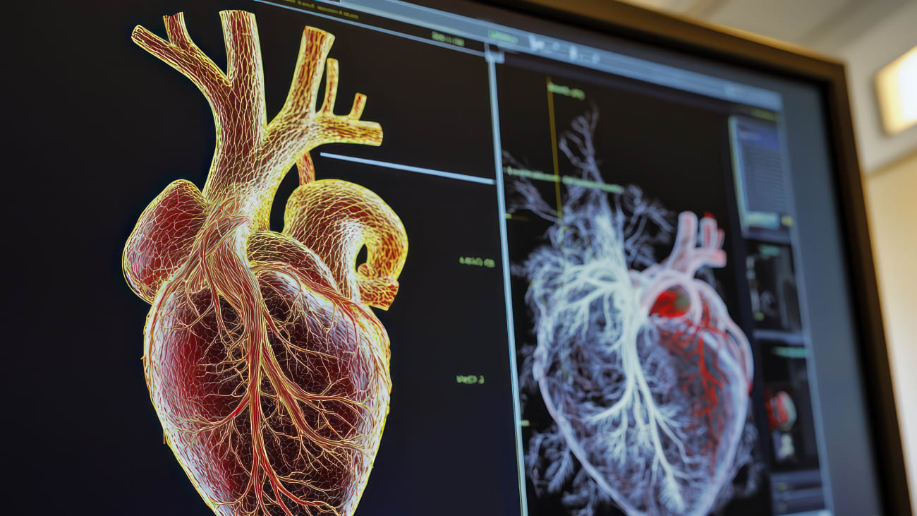
Flurpiridaz F-18 is used during PET scans under rest or stress conditions to identify the presence of CAD, myocardial viability (distinguishing dead from hibernating heart tissue) and microvascular dysfunction.
“Offering flurpiridaz F-18, a PET myocardial perfusion imaging (MPI) radiotracer, is a significant clinical and technological milestone. It provides superior sensitivity and higher image resolution, which translates into a more accurate diagnosis. We are excited to offer this to our patients,” said Socrates Kakoulides, M.D., a cardiologist with the Institute who championed the project.
“The new PET MPI radiotracer is used to assess blood flow to the heart muscle and its higher diagnostic accuracy can be beneficial, particularly in women, obese patients and those who are suspected to have multi-vessel disease,” said Hao Vuong, M.D., director of nuclear medicine at Baptist Health Baptist Hospital.
Flurpiridaz F-18 is the first new perfusion radiopharmaceutical in nearly three decades. The radiopharmaceutical agent has a longer half-life than existing PET MPI tracers, making it more accessible and eliminating the need for on-site tracer manufacturing.
CAD is the most common form of heart disease and is characterized by plaque buildup in the arteries. It is the leading cause of death for men and women in the U.S., according to the Centers for Disease Control and Prevention.
“We are at the forefront of cardiovascular imaging innovation, offering diagnostic capabilities not yet available elsewhere,” said Ricardo Cury, M.D., medical director of cardiac imaging for Miami Cardiac & Vascular Institute and Baptist Hospital. “To increase survival rates from cardiovascular events and to improve treatment, it’s important to provide an early and accurate diagnosis. This technology is a step forward.”
The initiative was launched through the collaboration of the Institute’s Imaging team championed by Drs. Kakoulides, Vuong and Cury.